

Historical profile. Every peptide medicine, from insulin analogs to semaglutide, depends on a simple chemical fact: the order of amino acids matters. Change that order and the molecule’s properties may change with it.
Today, researchers can read protein sequences, manufacture peptides and deliberately modify individual amino acids. In the 1940s, however, no one had yet determined the complete sequence of a protein.
Frederick Sanger, a Cambridge biochemist, helped change that by spending roughly 12 years working out the order of insulin’s 51 amino acids. It was an extraordinary exercise in chemical patience, involving fragmented proteins, filter paper, electric currents and the reconstruction of molecular information one piece at a time.
The achievement won him the 1958 Nobel Prize in Chemistry.
Then he turned to an entirely different molecular alphabet, developed methods for reading DNA and won a second Nobel Prize in 1980.
He remains the only person to have won the Nobel Prize in Chemistry twice.
Frederick Sanger, a Quaker Chemist in Wartime Cambridge
Frederick Sanger was born on August 13, 1918, in Rendcomb, Gloucestershire, England, into a family shaped by Quaker beliefs and a strong interest in science.
His father was a physician, and medicine initially seemed a natural career. But Sanger became more interested in understanding biological processes through experimental chemistry.
He entered St John’s College, Cambridge, studied natural sciences and completed his undergraduate degree in 1939.
When the Second World War began, Sanger registered as a conscientious objector. Influenced by the pacifist principles of his upbringing, he remained in Cambridge and continued his scientific education.
He completed his PhD in 1943 under Albert Neuberger, studying the metabolism of the amino acid lysine.
Afterward, Sanger began working with Charles Chibnall, whose research group had been investigating the chemistry of proteins, including insulin.
Insulin was an attractive research target for practical reasons.
It had been used to treat diabetes since the 1920s, was available in relatively pure form and was small compared with many other proteins.
But small was relative.
Insulin contains 51 amino-acid residues arranged in two chains. Determining the complete order of those residues was far beyond the routine analytical methods available at the time.
Scientists knew that proteins contained amino acids. What remained uncertain was whether a particular protein possessed one defined sequence and how that sequence could be established experimentally.
Sanger approached the problem as a chemist.
Rather than trying to examine the entire molecule at once, he would find ways to break it into smaller pieces, identify their contents and reconstruct the original order.
The strategy sounds straightforward today. Developing methods capable of making it work occupied much of the next decade.
Tagging the End of the Chain
Sanger’s first breakthrough involved a chemical reagent called 2,4-dinitrofluorobenzene, often abbreviated DNFB and later widely known as Sanger’s reagent.
The reagent reacts with free amino groups, allowing researchers to label the amino-terminal end of a protein chain.
After breaking the protein into smaller components, the labeled amino acid could be isolated and identified.
This gave Sanger a way to establish where a chain began.
But identifying the beginning was only the first step. The much harder problem was determining the sequence of the remaining amino acids.
Sanger used chemical and enzymatic methods to break insulin into shorter peptides.
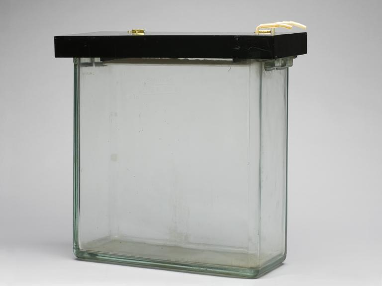
Those fragments could then be separated using techniques including paper electrophoresis and chromatography.
In electrophoresis, an electric field causes charged molecules to move through a supporting medium at different rates. Chromatography separates molecules according to their differing interactions with the materials through which they travel.
Combining these approaches helped produce distinct patterns of peptide fragments on filter paper.
The resulting arrangement of separated spots became known as a peptide fingerprint.
Each fragment contained part of the original sequence.
By determining the amino-acid order within different fragments and comparing overlapping sections, Sanger and his colleagues could gradually reconstruct longer chains.
The process resembled assembling a complicated puzzle whose pieces had no printed picture.
One fragment might establish a short stretch of amino acids. Another, overlapping fragment could reveal which residues came before or after it.
Gradually, the complete order emerged.
It was not the work of one person operating in isolation.
Important papers from the project included contributions by E. O. P. Thompson, L. F. Smith, A. P. Ryle and R. Kitai, among other collaborators.
In 1953, Sanger and Thompson published detailed studies of the amino-acid sequence of insulin’s glycyl chain.
Further work established the structure of the two chains and the disulfide bonds connecting them.
Preliminary findings on the disulfide bridges appeared in 1954, followed by a substantial 1955 publication examining the bonds linking the insulin chains.
The completed structure contained 51 amino acids arranged in two polypeptide chains, connected by two disulfide bonds, with a third disulfide bond within one chain.
According to the MRC Laboratory of Molecular Biology, the work involved approximately 12 years of dedicated research.
He broke insulin into pieces, separated them on paper and reconstructed a molecular sequence that no one had previously been able to read.
Why the Sequence Changed Everything
Determining insulin’s sequence settled a fundamental question about protein chemistry.
It established that a protein could possess a precise and reproducible primary structure, rather than being an irregular collection of amino acids whose arrangement varied unpredictably.
That discovery changed how scientists understood the relationship between chemical structure and biological function.
Amino-acid order helps determine how a protein folds, interacts with other molecules and performs its biological role.
Sanger’s insulin work did not, by itself, explain every aspect of protein folding or three-dimensional structure. Those questions required additional discoveries and experimental methods.
But it established that the primary structure could be determined.
This gave biochemists a foundation for studying other proteins with the same chemical logic.
In 1958, the Nobel Committee awarded Frederick Sanger the Nobel Prize in Chemistry for his work on protein structure, especially insulin.
He received the prize alone, even though the experimental project had included important contributions from colleagues.
The achievement became one of the foundations of modern molecular biology.
It also supplied essential knowledge for developments in therapeutic protein engineering.
Knowing insulin’s amino-acid order was distinct from knowing its three-dimensional structure. The latter became an important part of the research led by Dorothy Hodgkin, whose crystallographic work Grey Pepper explored in its profile of Hodgkin and insulin.
Later, recombinant-DNA technology enabled researchers to manufacture human insulin using engineered microorganisms, a development discussed in our profile of Axel Ullrich and recombinant insulin.
Today, peptide drug developers routinely investigate how changing particular amino acids, introducing structural modifications or altering molecular attachments affects receptor activity, stability and duration of action.
That work depends on knowing exactly which amino acids are present and how they are arranged.
Modern peptide engineering assumes that amino-acid sequences can be identified and deliberately modified. Sanger helped establish the scientific foundation for that work.
The Second Nobel: From Proteins to DNA
Sanger did not spend the rest of his career sequencing proteins.
In 1962, he joined the newly established Medical Research Council Laboratory of Molecular Biology in Cambridge, where he became Head of the Protein and Nucleic Acid Chemistry Division.
He increasingly turned his attention to nucleic acids, beginning with RNA and eventually moving to DNA.
DNA presented a related analytical challenge.
Instead of determining the order of amino acids, scientists needed to establish the precise order of four nucleotide bases.
Those bases are commonly represented by the letters A, T, G and C.
With Alan Coulson, Sanger developed the plus-and-minus method for determining DNA sequences in the mid-1970s.
This technique helped enable one of the most important early milestones in genomics: the sequencing of the bacteriophage phiX174 genome.
In 1977, Sanger and colleagues reported a provisional sequence of the viral genome, which contains 5,386 nucleotides.
The following year, a published analysis described completion and confirmation of the sequence using improved methods, with revisions to the earlier provisional findings.
It was the first complete DNA genome sequence.
Meanwhile, Sanger, Steve Nicklen and Alan Coulson developed a more efficient sequencing technique based on terminating DNA chain extension at specific nucleotides.
The method, described in a landmark 1977 paper in the Proceedings of the National Academy of Sciences, used nucleotide analogues that stopped DNA polymerase from extending a growing DNA strand.
By examining fragments of different lengths, researchers could infer the sequence of the original DNA.
This became known as dideoxy chain-termination sequencing, or simply Sanger sequencing.
It was faster and more accurate than the earlier plus-and-minus approach, and it became a fundamental technique in molecular genetics.
In 1980, Sanger received his second Nobel Prize in Chemistry.
The prize was divided between three scientists. Paul Berg received half for work involving recombinant DNA, while Walter Gilbert and Frederick Sanger each received one-quarter for contributions to determining nucleic-acid base sequences.
Sanger’s achievement was exceptional.
He had helped establish how to read the amino-acid sequence of proteins, then helped develop the technology for reading DNA.
Both achievements changed the scale of questions that biologists could investigate.
He retired in 1983. During the following decade, a new large-scale genome research center was named in his honor, later becoming the Wellcome Sanger Institute.
His DNA sequencing methods became part of the technological foundation for the Human Genome Project and the genomics revolution that followed.
What the Legend Leaves Out
Stories about scientific genius often compress years of difficult experimental work into a single discovery.
Sanger’s career resists that simplification.
The insulin project took roughly 12 years, required the development and refinement of analytical techniques, and depended on the contributions of multiple researchers.
The later DNA sequencing breakthroughs were collaborative too.
Alan Coulson, Steve Nicklen and other colleagues made substantial contributions to the methods and experiments associated with the second Nobel Prize.
That does not diminish Sanger’s achievement. It makes the history more accurate.
The scientific progress came from sustained experimental work, not simply from a flash of insight.
By contemporary accounts, Sanger was also notably modest.
Colleagues remembered a scientist who preferred the laboratory bench to administration, public attention or grand pronouncements about his own importance.
In the University of Cambridge’s 2013 obituary, Richard Henderson recalled a young researcher asking Sanger for advice and receiving a short answer: “I think you should try harder.”
It was an unusually plain piece of advice from someone whose discoveries required remarkable persistence.
Sanger died on November 19, 2013, at the age of 95.
He had lived to see protein sequencing become an established scientific discipline, DNA sequencing become the foundation of genomics, and insulin become one of many protein and peptide medicines whose molecular structures could be deliberately engineered.
The connection between his two Nobel Prizes is the deepest part of the story.
First, he helped demonstrate that the chemical sequence of a protein could be read.
Then he helped create a way to read the genetic material that encodes those sequences.
The first accomplishment changed protein chemistry. The second transformed genetics.
Modern molecular biology runs on the assumption that life’s chemical instructions can be identified, read and analyzed. Frederick Sanger helped turn that assumption into an experimental reality—twice.
Sources and Further Reading
- NobelPrize.org. Frederick Sanger: Biographical. Nobel Prize in Chemistry 1958.
- Royal Swedish Academy of Sciences. Nobel Prize in Chemistry 1980: Press Release. October 14, 1980.
- MRC Laboratory of Molecular Biology. 1958 Nobel Prize in Chemistry: Fred Sanger and Protein Sequencing.
- University of Cambridge, Department of Biochemistry. Frederick Sanger: Research and Nobel Prizes.
- Sanger F, Thompson EOP. The Amino-Acid Sequence in the Glycyl Chain of Insulin. I. The Identification of Lower Peptides From Partial Hydrolysates. Biochemical Journal, 1953.
- Ryle AP, Sanger F, Smith LF, Kitai R. The Disulphide Bonds of Insulin. Biochemical Journal, 1955.
- Sanger F, Nicklen S, Coulson AR. DNA Sequencing With Chain-Terminating Inhibitors. Proceedings of the National Academy of Sciences, 1977.
- Sanger F, et al. The Nucleotide Sequence of Bacteriophage phiX174. Journal of Molecular Biology, 1978.
- University of Cambridge. Dr Frederick Sanger (1918–2013). November 20, 2013.
- Science Museum Group Collection. Sanger’s Electrophoresis Equipment, Object 1988-206.