

Insulin is usually discussed as a pancreas-and-liver hormone, the 51-amino-acid peptide that helps regulate blood sugar. But the brain responds to insulin too, and researchers have spent decades investigating whether disrupted brain insulin signaling helps connect metabolic disease with cognitive decline.
The problem is measuring that signaling in a living person. A small randomized trial published in Food & Function offers an unusual approach: researchers administered insulin through the noses of older volunteers, measured cerebral blood flow using MRI, and asked whether eating peanuts every day changed the response.
The result was intriguing but narrow. After 16 weeks of daily peanuts, one region of the right brain showed a significantly smaller blood-flow response to intranasal insulin than after a control period. Most other measures did not change. The researchers interpreted the regional finding as potentially beneficial, but the experiment did not establish that peanuts improve brain insulin sensitivity, prevent memory loss or protect against dementia.
The distinction matters. This was a study of 31 generally healthy older adults and an indirect imaging measure, not a clinical trial demonstrating the prevention or treatment of neurological disease.
How Do You Measure Brain Insulin Resistance?
Insulin has important functions outside the bloodstream’s familiar glucose-regulation story. In the brain, it participates in pathways involved in neuronal activity, appetite, energy balance and cognition. Researchers are particularly interested in how brain insulin responses change with aging, obesity and metabolic disease.
But measuring those responses directly is difficult. Brain tissue cannot be routinely sampled from healthy volunteers, and an ordinary blood test does not reveal how individual brain regions react to insulin.
Intranasal insulin offers an experimental workaround. Researchers administer the peptide through the nose to investigate central nervous system responses while limiting some of the systemic effects associated with conventional administration. The nasal route has attracted attention for other peptide-based treatments, including experimental GLP-1 nasal delivery approaches.
In this study, participants received 160 international units of insulin intranasally. Researchers measured cerebral blood flow before administration and again approximately 30 minutes afterward.
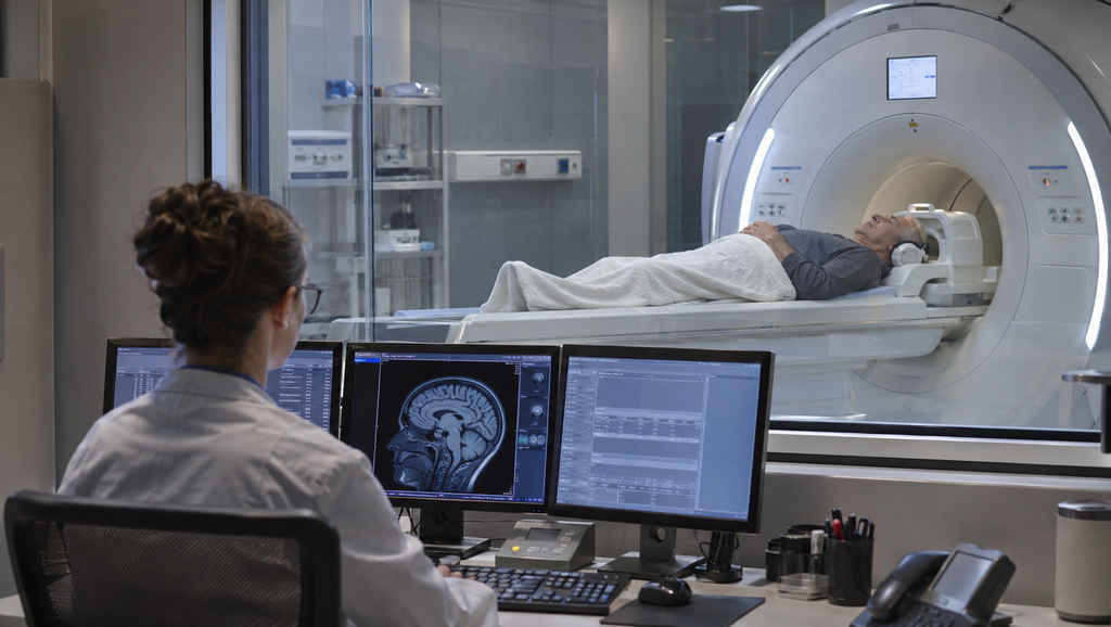
The imaging method was arterial spin labeling magnetic resonance imaging, or ASL-MRI. It uses magnetically labeled blood water to estimate tissue perfusion without requiring an injected contrast agent.
The difference in cerebral blood flow before and after the insulin challenge served as an indirect marker of brain insulin responsiveness. The researchers could examine whether different dietary conditions were associated with different regional blood-flow responses.
There is an important limitation built into the technique. Cerebral blood flow can reflect changes in neuronal and vascular activity, but it does not measure insulin-receptor activation directly. A smaller blood-flow response is not automatically evidence of healthier neurons or more efficient insulin signaling.
The study measured blood flow, not neurons. That distinction is central to understanding the result.
The Peanut Experiment: 31 Older Adults, Two Diets, 16 Weeks Each
The study was conducted by researchers at Maastricht University Medical Center in the Netherlands. It used a randomized, single-blind, controlled crossover design in generally healthy older adults.
Thirty-six participants were randomized and 31 completed the trial. The completers included 15 women and 16 men, with an average age of 67 years and an average body mass index of 26.7.
Each participant completed two 16-week study periods separated by an eight-week washout. During one period, volunteers consumed 60 grams of unsalted, skin-roasted peanuts daily, provided in prepackaged portions. During the control period, they avoided peanuts and other nuts.
The order was randomized. Some participants began with peanuts, while others began with the control condition. After the washout, they switched to the other intervention.
That crossover design was valuable because each person could serve as their own comparison. Differences between individuals, including age, physiology and habitual diet, therefore had less opportunity to distort the intervention comparison than in a simple observational study.
It was not, however, a calorie-matched experiment. The control group did not receive a substitute snack matching the peanut portions for energy or nutrients.
During the peanut period, reported calorie intake increased, and participants consumed more unsaturated fat and fiber while their intake of carbohydrates and saturated fat shifted downward. These changes matter because they make it difficult to identify whether any observed biological effect came from peanuts themselves, a particular nutrient, or the overall change in dietary composition.
At the end of each intervention period, the volunteers underwent the intranasal insulin challenge and MRI measurements. The investigators also examined blood markers, body composition and fat content in the liver.
One Brain Region Responded Differently After Peanuts
The clearest finding appeared in a localized region of the right cerebral hemisphere. Compared with the control period, the peanut period was associated with a smaller cerebral blood-flow response to intranasal insulin.
The reported mean difference was minus 5.4 milliliters per 100 grams of tissue per minute, with a reported variation of plus or minus 5.4. The identified cluster covered 552 cubic millimeters, and the statistical test yielded a P value of 0.044.
An anatomical atlas localized the cluster primarily to areas associated with the lingual gyrus, hippocampus and parahippocampal gyrus. These structures participate in visual processing and aspects of memory, although their functions are more complex than any single label suggests.
The researchers described the regional response as potentially beneficial. Their interpretation rests partly on the complex relationship between insulin, neuronal signaling and blood flow in different brain regions. In some circumstances, insulin-related inhibitory signaling could be associated with reduced neuronal activity and a smaller vascular response.
That explanation remains a hypothesis rather than a directly measured result. The experiment did not demonstrate that the smaller blood-flow response represented improved insulin-receptor function, healthier brain tissue or a reduced risk of cognitive decline.
It is also important not to confuse a change in the insulin-induced response with a decline in ordinary resting cerebral blood flow. They are different physiological measurements.
No other significant clusters emerged from the whole-brain analysis. Across broader brain compartments, including global, cortical, gray-matter and subcortical measurements, the investigators did not observe statistically significant differences in insulin-induced blood-flow responses between the two dietary conditions.
One regional response changed. The rest of the brain did not show a comparable statistically significant signal.
What Did Not Change, and What the Memory Results Cannot Prove
The list of metabolic outcomes without statistically significant differences was considerably longer than the list of positive findings.
Fasting blood glucose, fasting insulin and HOMA-IR, a commonly used estimate of whole-body insulin resistance, did not differ significantly between the peanut and control periods. The researchers also found no significant difference in liver fat content.
Body fat percentage and waist circumference remained broadly stable despite the reported increase in energy intake during the peanut intervention. Total cholesterol, LDL cholesterol and HDL cholesterol were not significantly altered.
Triglycerides were lower at week eight, but that difference was not sustained at week sixteen. The lipid analyses were exploratory and were not adjusted for multiple outcome comparisons, making isolated significant results less persuasive.
High-sensitivity C-reactive protein, a marker of systemic inflammation, also did not change significantly.
There was an earlier cognitive finding from the same participants. In a separate paper published in Clinical Nutrition in 2025, the investigators reported that peanut consumption increased global resting cerebral blood flow by 3.6% and improved performance by 5.8% on a delayed verbal-recognition memory measure.
That earlier study also reported a reduction in systolic blood pressure. But it did not establish that peanuts prevent dementia or produce generalized cognitive improvements across all tested domains.
The new insulin-response analysis explored whether its regional imaging findings could help explain those previously observed memory changes.
The researchers found no statistically significant correlation between the change in regional brain insulin responsiveness and the earlier improvement in verbal memory.
They cautioned that the sample was not designed to establish such a relationship. The absence of a significant association therefore does not prove the mechanisms are unrelated, but neither does it support a claim that the insulin response caused the memory improvement.
The same 31 people contributed to both analyses. Two papers describing related findings from one experiment should not be mistaken for two independent clinical confirmations.
Calories went up, body fat did not, and one brain region behaved differently. That is a scientific lead, not a verdict.
Why the Direction of the Insulin Response Is Hard to Interpret
One of the most interesting aspects of this research is also its central interpretive difficulty. A smaller blood-flow response after an insulin challenge could reflect a change in how the brain regulates neuronal or vascular activity. It does not necessarily establish that tissue has become more sensitive to the hormone.
The meaning of insulin-induced blood-flow changes varies across brain regions, and the relationship among receptor signaling, neuronal inhibition, energy metabolism and perfusion remains incompletely understood.
Consequently, identifying a statistically significant regional difference is not equivalent to identifying a clinically meaningful improvement. The regional P value of 0.044 supports a finding under the researchers’ statistical model, but the small sample and localized effect limit how confidently it can be generalized.
The dietary design creates another uncertainty. Without an energy- and nutrient-matched control food, the investigators cannot isolate an effect unique to peanuts.
They discuss several plausible biological contributors, including L-arginine, which participates in nitric oxide production, and the unsaturated fats, fiber and polyphenols found in peanuts. Nitric oxide is involved in vascular signaling and could plausibly influence cerebral perfusion.
But the study did not directly compare those components or establish their individual contributions to the observed MRI response.
The population was another limitation. Participants were generally healthy older adults, rather than people with diabetes, diagnosed cognitive impairment or Alzheimer’s disease. A result in this group cannot be assumed to apply to patients with established metabolic or neurological disorders.
Peanut consumption was generally well tolerated in the trial. One participant withdrew because of gastrointestinal discomfort following peanut intake, and the researchers reported no study-related serious adverse events. That does not remove the established allergy risk associated with peanuts or turn the intervention into a recommendation for every older adult.
The research also received funding from The Peanut Institute Foundation. According to the publication’s funding disclosure, the foundation did not participate in the study design, acquisition or analysis of data, publication decisions or manuscript preparation. This commercial-interest context is relevant when interpreting nutrition research, although funding alone does not determine whether a result is scientifically valid.
Why This Small Trial Matters Beyond Peanuts
The most interesting contribution may not be the food. It may be the method.
Many studies of nutrition and brain health rely on cognitive tests, questionnaires or broad measurements of metabolic health. Those outcomes can be useful, but changes may be difficult to detect over relatively short periods, and they rarely reveal what is happening in a specific brain region.
Combining a peptide challenge with advanced MRI offers another way to investigate the biology. Researchers can ask whether dietary interventions, exercise programs or experimental medicines change the way particular brain regions respond to a hormone.
The broader history of peptide research depends on techniques that make previously inaccessible physiology measurable. Rosalyn Yalow and Solomon Berson’s work on measuring insulin helped transform hormone research decades ago. Modern imaging methods pose a related question: not simply how much hormone is present, but how different tissues respond to it.
That question is increasingly relevant as GLP-1 drugs and other metabolic treatments are investigated for possible effects on brain function. Researchers need methods capable of distinguishing central nervous system effects from changes associated with weight loss, blood sugar or cardiovascular physiology.
Intranasal insulin challenges and regional perfusion imaging may contribute to those investigations. But they must be validated carefully before an imaging response can serve as a dependable indicator of clinical benefit.
The next useful peanut study would involve a larger and more diverse population, a calorie- and nutrient-matched comparison, predefined imaging outcomes and cognitive assessments capable of determining whether the biological changes correspond to meaningful functional improvements.
For now, this trial provides no evidence that daily peanuts prevent dementia, reverse diagnosed brain insulin resistance or substitute for established medical care. It describes a measurable difference in one regional response following 16 weeks of dietary intervention.
The result deserves follow-up because the method is intriguing and the biological question matters. It does not deserve to be marketed as a proven brain-health treatment.
Thirty-one people ate peanuts, researchers challenged their brains with insulin, and one localized blood-flow response changed. Understanding what that change means is the next study, not the conclusion of this one.
Research funding disclosure: The study was funded by The Peanut Institute Foundation. The authors reported that the funder had no role in study design, data collection or analysis, publication decisions or preparation of the manuscript.
Sources
- Kerkhof L, Mensink RP, Plat J, Nijssen KMR, Joris PJ. Daily consumption of unsalted, skin-roasted peanuts beneficially affects regional brain insulin responsiveness in older adults: results of a longer-term randomized controlled trial. Food & Function. 2026;17(20):9354–9364. DOI: 10.1039/D6FO03208G.
- Kerkhof L, et al. Longer-term skin-roasted peanut consumption improves brain vascular function and memory: A randomized, single-blind, controlled crossover trial in healthy older adults. Clinical Nutrition. 2025;55:170–179.
- Maastricht University. Institutional publication record for the 2026 brain insulin responsiveness study, including research funding disclosure.
- News-Medical. Eating peanuts every day may change your brain’s insulin response. November 4, 2026.
- ClinicalTrials.gov. NCT05724654 — Registered peanut intervention trial.